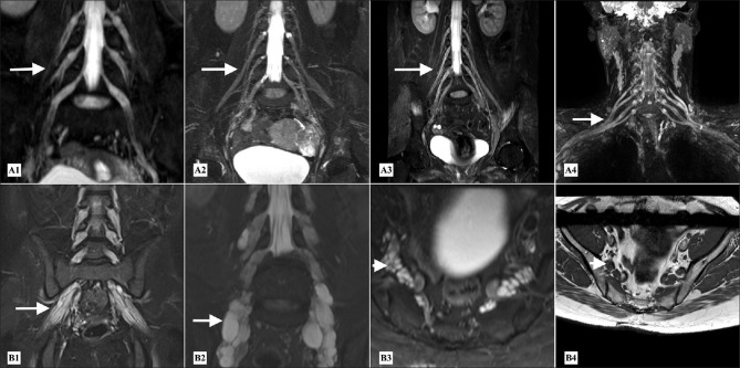
Figure 6.
CIDP - MR Neurography (A1-A3) STIR MIP coronal reconstructed images of the lumbosacral plexuses in three different patients and (A4) of the brachial plexus, showing mild to moderate uniform thickening of the roots and branches of the lumbosacral plexuses (A1-A3) (arrows) and of the roots and trunks of brachial plexus (A4) (arrow) in acquired demyelinating neuropathies. CMT - MR Neurography (B1 and B2) Coronal STIR MIP reconstructed images of the lumbosacral plexus and (B3- B4) STIR axial and T1 axial images at the level of the lumbosacral plexus showing thickening of the roots and proximal branches of lumbosacral plexuses (arrows). There is increased inter-fascicular epineural fat with preserved fascicles (B3-B4) (arrowheads) giving multi cystic appearance of the thickened nerve roots in inherited neuropathies

