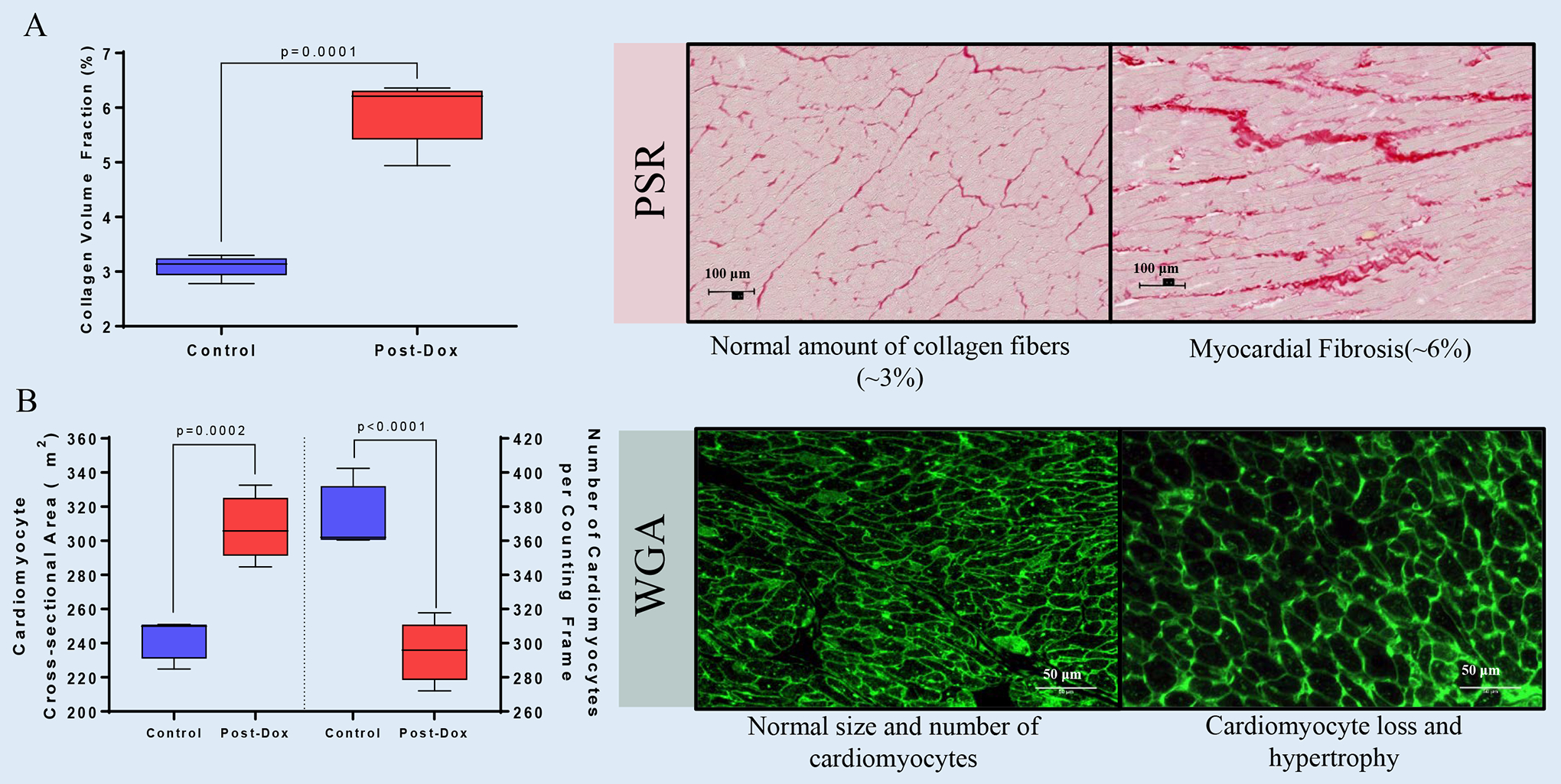
Figure 1. Histopathological Mechanisms Leading to CMR-derived Extracellular Volume Fraction Expansion after Chronic Receipt of Doxorubicin.

(A) Graphical representation of cardiac fibrosis and (B) cardiomyocyte size and numbers and representative microphotographs. All values are mean ± SEM at baseline.
