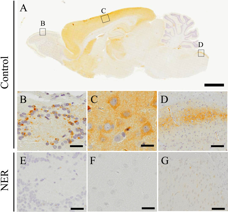
Fig. 1.
Immunohistochemistry for PHF24. PHF24 Immunohistochemistry on sagittal section of the CNS in the control rats (A). Black squares indicate the areas of B–D. The expression of PHF24 is mainly observed in the periglomerular layer (B), cerebral cortex (C) and medulla oblongata (D). While PHF24 is expressed broadly in the cerebral cortex (C), the characteristic expression of PHF24 is found in the periglomerular layer (B) and medulla oblongata (C). In the NER, prominent decreased expression is found in the CNS; periglomerular layer (E), cerebral cortex (F) and medulla oblongata (G). Bars: 2 mm (A), 20 µm (B–G).

