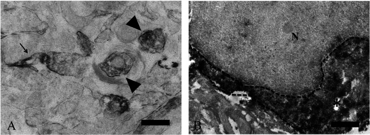
Fig. 4.
Immunoelectron microscopy for PHF24 in the spinal cord and olfactory bulb. Expression of PHF24 located in presynaptic terminals (A: arrowheads) and synaptic membrane (A: arrow). This finding is observed both in the spinal cord and olfactory bulb and a representative example in the spinal cord is presented (A). In the olfactory bulb, positive reaction for PHF24 is diffusely found in the neuronal cytoplasm (B: asterisk). N: Nucleus Broken line: a border of cytoplasm Black bars: 100 µm (A), 1 µm (A, B).

