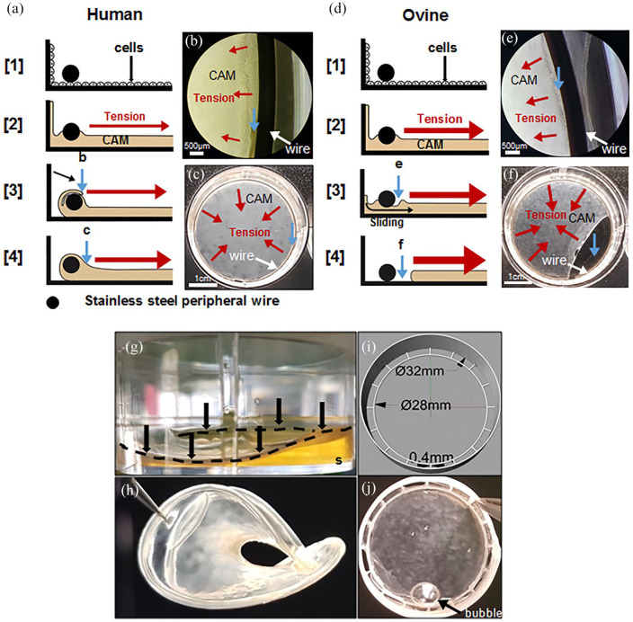
Figure 8.
Ovine CAM sheets often detached from stainless steel wire anchors but strongly attached to PLA anchors. (a–f) montage illustrating the difference between the development of human and ovine sheet attachment to the steel peripheral wire anchor. Cells grew to confluence under the wire (step [1]) and then started depositing the ECM to form the CAM sheet (step [2]). Red arrows show the direction of the force generated by tissue tension. (a) Human cells grew on and CAM accumulated near the wire slowly enveloping it, meanwhile, the CAM detached from the wall and folded over the wire enveloping it (steps [3] and [4]). (b) Phase-contrast micrograph showing folding over the anchor (at 3 weeks). (c) Macroscopic view of a well-attached human CAM sheet at the bottom of a 6-well plate (at 8 weeks). Blue arrows with lowercase letters show the same region shown by the blue arrows of the matching lowercase figure. White arrows point to the anchor. (d) Ovine cells were more contractile and developed earlier/larger tissue tension pulling away the CAM accumulating against the wire before it could envelop it (step [3]). Without a strong attachment, the sheets slipped under the wire and contracted (step [4]). (e) Phase-contrast micrograph of an ovine sheet showing tissue separation from the anchor and a lack of folding after 1 week. (f) Macroscopic view showing sheet detachment a week later. (g) Ovine sheets produced with a PLA peripheral wire anchor (black arrows and discontinued black line) in a 6-well plate after 3 weeks. PLA anchors allow strong attachment of ovine sheets but tissue tension was strong enough to bend the PLA leading to the lifting of the sheet from the culture substrate (s). (h) After 4 weeks of culture, the structure is completely folded and unusable. (i and j) PLA anchorage devices were 3D-printed with a reinforced outer ring connected to an inner wire allowing strong ovine sheet attachments and withstood tissue contractile forces. A medium bubble can be seen on the sheet (black arrow).

