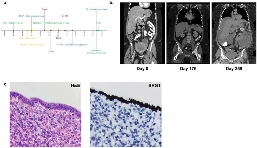
Figure 2: Clinical Summary of Patient 2 diagnosed with SMARCA4-DUS.
(a) Timeline of major clinical events. (b) Computed tomography (CT) scans at initial presentation (left panel), showing no evidence of disease (NED) following completion of 4 cycles of AIM chemotherapy (middle panel), and after re-presentation with diffuse metastatic disease (right panel). (c) Pathological evaluation with H&E staining revealing rhabdoid morphology and sheets of highly mitotically active cells characteristic of SMARCA4-DUS (left panel), with immunohistochemistry demonstrating loss of BRG1 (right panel).

