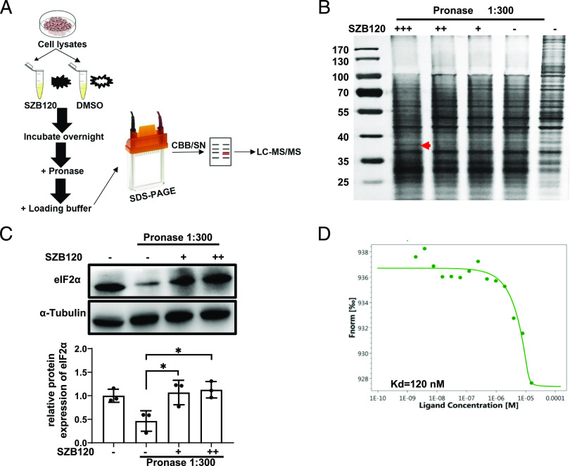
FIGURE 2.
Identification of eIF2α as a direct target protein of SZB120 by DARTS. (A) The schematic illustration of the DARTS technique. (B) The cell lysates were treated with DMSO or SZB120 (1, 10, 100 μM) overnight at 4°C, followed by SDS-PAGE analysis and Silver Staining. The red arrow indicates a protective band by SZB120 of ∼38 kDa. (C) DARTS and Western blotting to confirm the potential binding target eIF2α of SZB120 (10 and 100 μM). The lower panel is the quantitative results. Data are shown as a fold of α-tubulin (n = 3). (D) MST analysis of the binding affinity between SZB120 and eIF2α. The fitted Kd value is shown. *p < 0.05.

