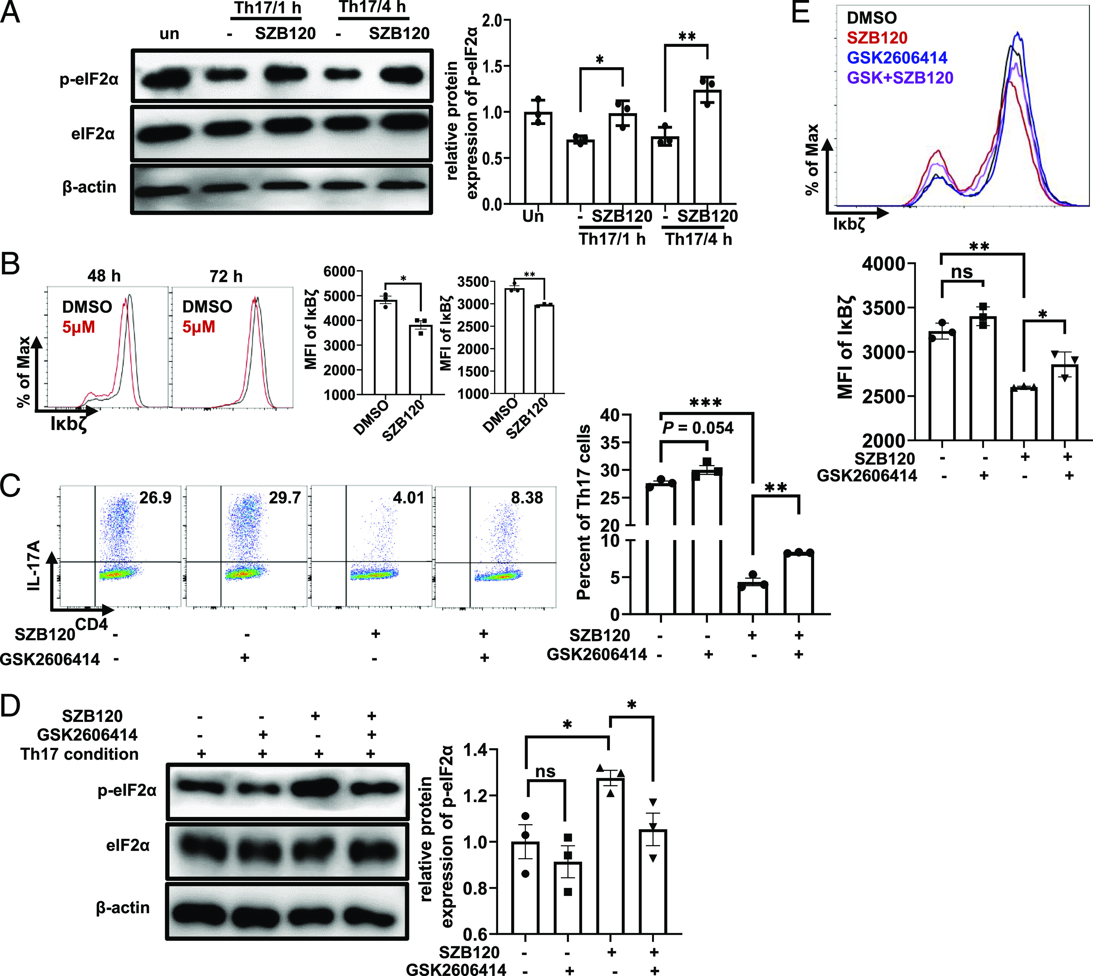
FIGURE 3.

SZB120 inhibits IL-17A secretion in an eIF2α phosphorylation-dependent manner. (A) Unstimulated or TCR-activated Th17 cells were treated with DMSO and 5 μM SZB120 and lysed for Western blotting of p-eIF2α and eIF2α after 1 and 4 h. The right panel is the quantitative results. The data are shown as a fold of eIF2α (n = 3). (B) Naive CD4+ T cells were activated under Th17 cell polarization conditions and treated with DMSO and 5 μM SZB120. Cells were measured by flow cytometry to determine the IκBζ mean fluorescence intensity (MFI) of CD4+ T cells at the indicated times (n = 3). Data are the mean ± SEM by unpaired t tests. (C) T cells activated under Th17 polarizing conditions were treated with DMSO, SZB120 (5 μM), GSK2606414 (4 nM), or SZB120 plus GSK2606414 and stained for IL-17A intracellular cytokines (n = 3). (D) Unstimulated or TCR-activated Th17 cells were treated with the four groups as described in (C), and lysates were analyzed after 4 h by Western blotting of p-eIF2α and eIF2α. The quantitative results are shown in the right panel. Data are shown as a fold of eIF2α (n = 3). (E) Naive CD4+ T cells treated as described in (C) were differentiated under Th17 polarization conditions for 72 h. Cells were measured by flow cytometry to determine the IκBζ MFI of CD4+ T cells. The quantitative results are shown in the lower panel. All data represent at least three similar experiments with three samples per group in each. Data are the mean ± SEM by one-way ANOVA (A and C–E) with Bonferroni correction for multiple comparisons or the mean ± SEM by unpaired t tests (two-tailed) (B). *p < 0.05, **p < 0.01, ***p < 0.001.
