
Figure 1.
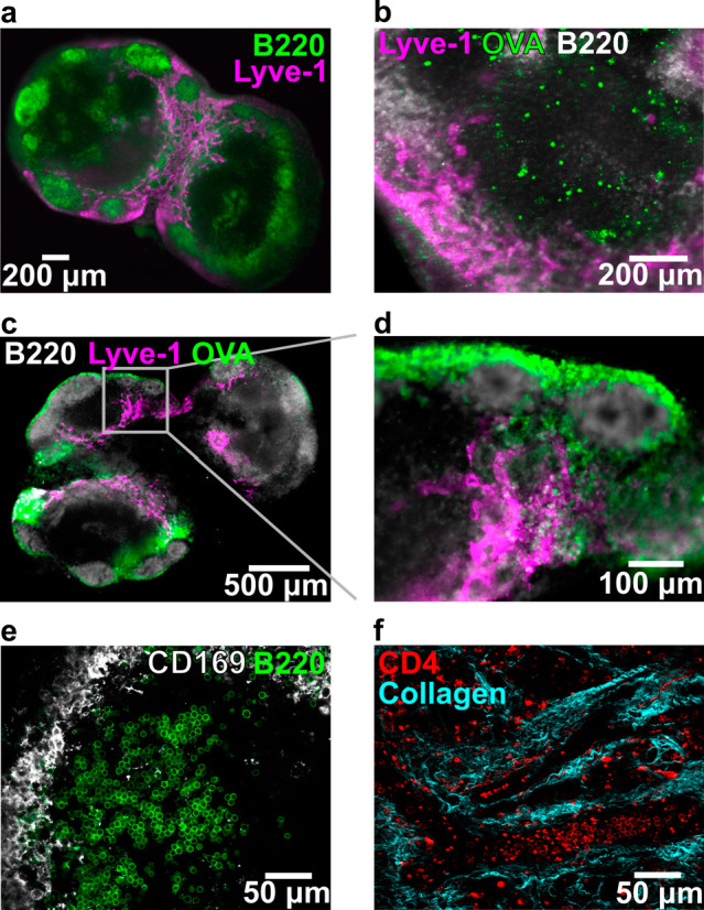
Key structural features remain intact in thick lymph node slices (iLN, aLN, bLN). (a) Slice labeled with anti-B220 (FITC, green) and anti-Lyve-1 (eFluor660, purple) revealed key structural features of the lymph node. Slice shown from a female C57Bl-6J mouse. (b–d) Slices from OVA (rhodamine labeled, green)-immunized C57Bl/6-J mice, labeled with anti-B220 (FITC, gray), and anti-Lyve-1 (eFluor660, purple). (b) Rhodamine-OVA was visible inside of cells within the T cell rich (B220-dim) region of the lymph node 3 days after immunization. (c) Rhodamine-OVA was visible in the sinuses and lymphatics 1 day after immunization; panel (d) shows inset. (e) High-definition image collected by confocal microscopy. Slice labeled with anti-B220 (AlexaFluor 647, green) and anti-CD169 (AlexaFluor 594, gray). (f) Image of lymph node slice collected by two-photon microscopy, showing CD4 positive T cells (FITC-CD4 Fab′) within the collagen matrix (second harmonic imaging). Detailed methods for each panel are provided in the SI.