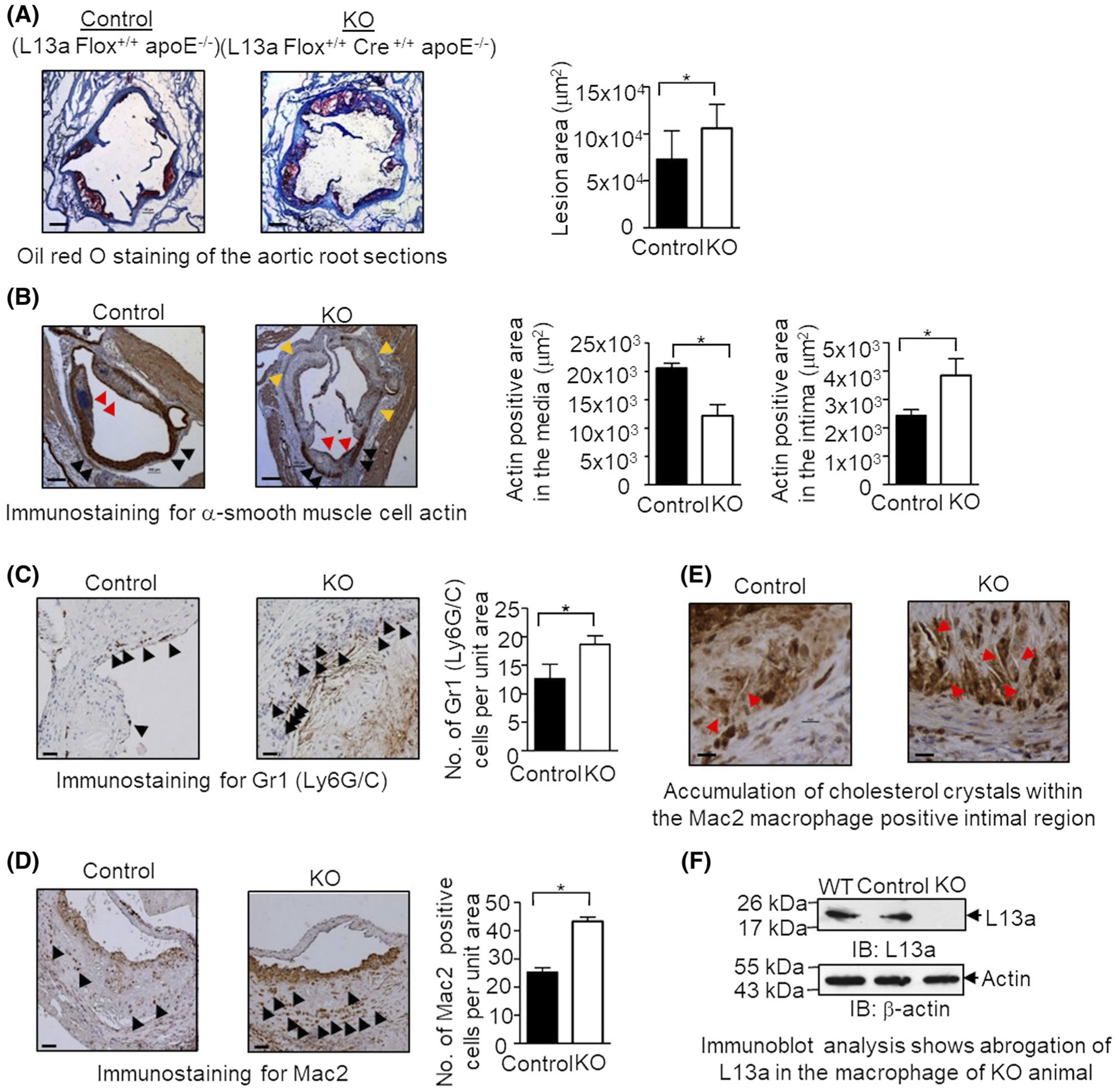
FIGURE 1.

High-fat diet (HFD)-induced atherosclerosis in myeloid cell-specific L13a deficient mice is associated with larger plaques and greater number of immune cells. A, Quantification of atherosclerotic lesion size of aortic roots in mice with or without macrophage-specific L13a deletion on ApoE−/− background and representative images of oil red O staining; Left panel: control group (L13a Flox+/+ApoE−/−), Right panel: KO group (L13a Flox+/+LysMcre+/+ApoE−/−). Mice were fed HFD for 14 weeks before euthanization. B-D, Immunohistochemistry of aortic plaques with α-smooth muscle cell actin antibody for SMC (B), RB6–8C5 antibody for neutrophils (C), and anti-Mac2 antibody for macrophages (D). Scale bars: 100 μm. In (B), black arrows point medial smooth muscle cell layer, red arrows point smooth muscle cells accumulated in the plaque and yellow arrows point areas of medial smooth muscle cell layer breakdown. Quantifications of each cell types are shown. E, Abundance of cholesterol crystals in control and KO mice groups observed in Mac2 staining. Scale bars: 100 μm. F, Confirmation of macrophage-specific depletion of L13a in KO mice. Peritoneal macrophage lysates were analyzed by immunoblot with anti-L13a antibody followed by the reprobing with anti-beta actin antibody. Results are expressed as mean ± SD, n = 4. *P ≤ .05
