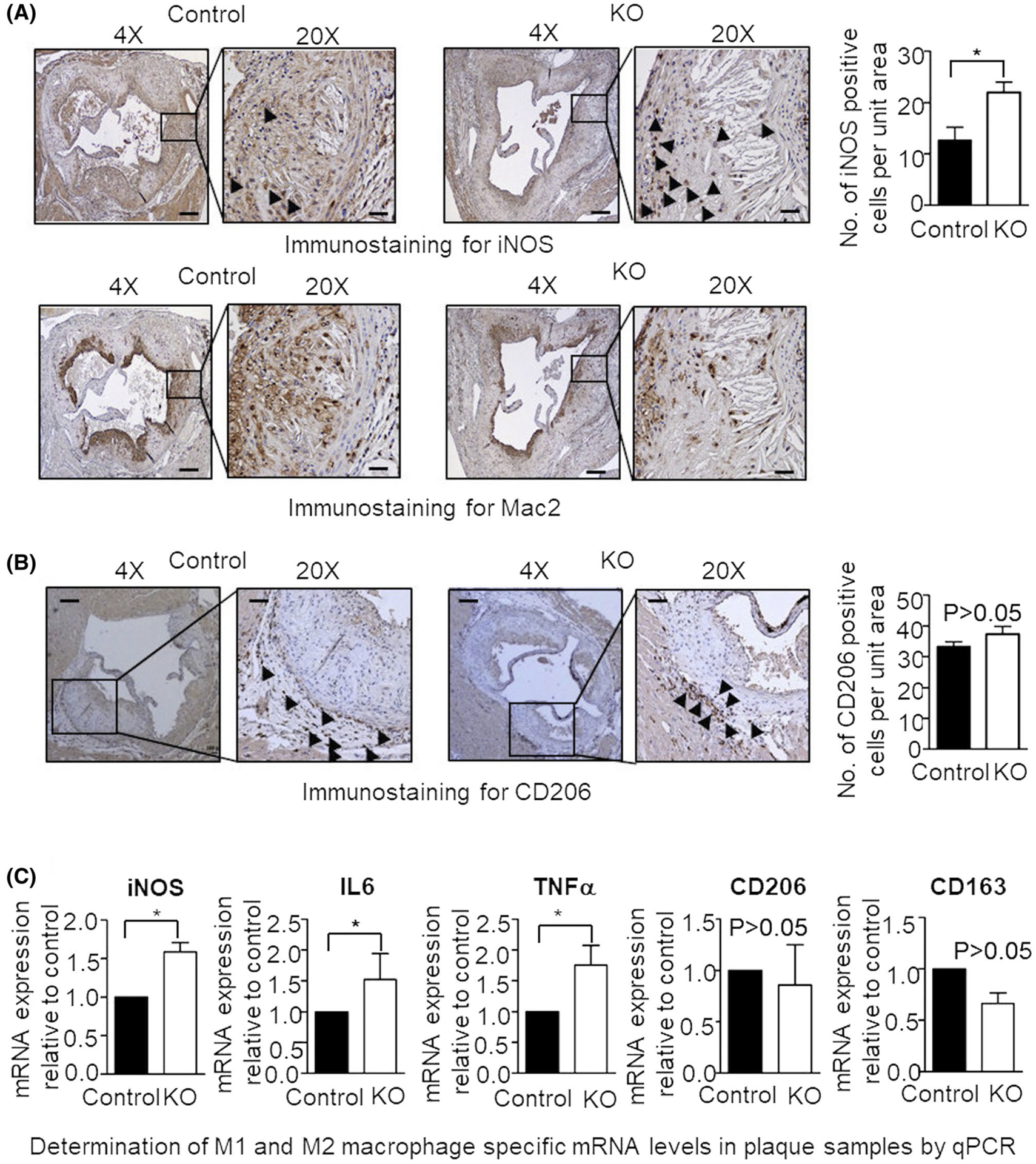
FIGURE 3.

HFD results in increased expression of M1 macrophage-specific markers in the atherosclerotic plaque of myeloid cell-specific L13a deficient mice. A and B, Immunohistochemical detection of iNOS (A), Mac2 (A) and CD206 (B) expressing cells in the aortic root sections prepared from control and KO mice. Quantifications for iNOS and CD206 are shown; Mac2 staining of the adjacent serial section shows the presence of macrophages in the same area. Scale bars: 100 μm. C, steady state levels of iNOS and other M1- and M2-type macrophage-specific mRNAs in aortic plaque cells were measured by real-time quantitative PCR. To generate cDNA template, mRNA obtained from laser microdissected cuts of aortic plaques were reverse transcribed. The levels of target mRNAs were normalized with mouse GAPDH mRNA and were presented as mean ± SD, n = 3. *P ≤ .05, compared with the control group
