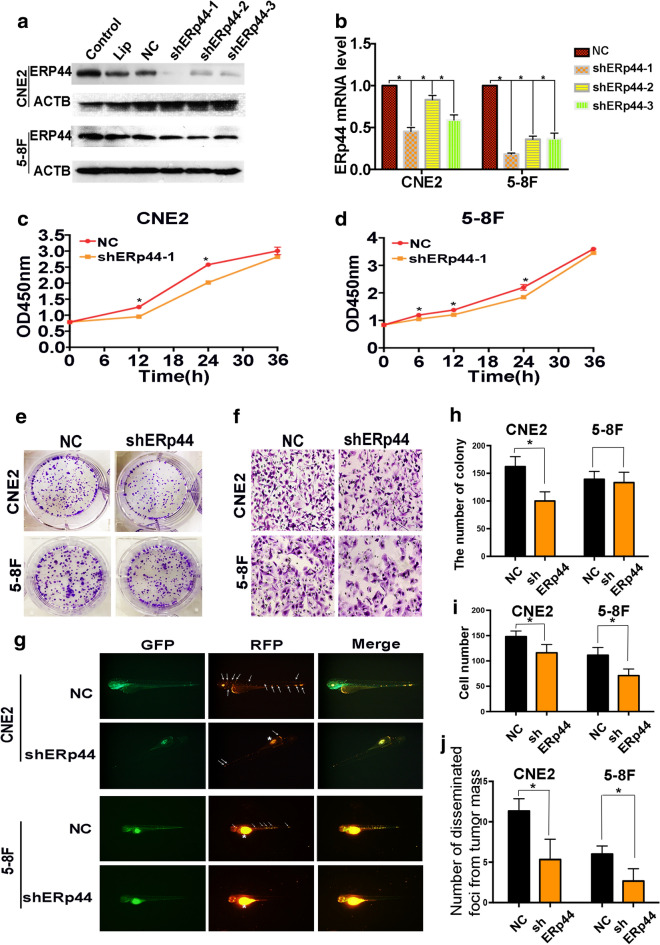
Fig. 2.
Interference of ERp44 expression could inhibit the malignant phenotype of NPC cells. a CNE2 and 5-8F cells were transfected with shRNA targeting ERp44 (sh-1, sh-2, sh-3) or a scrambled sequence (NC). b qRT-PCR was used to detect the mRNA level of ERp44 after the transfection. c, d CCK8 was used to determine cell proliferation after the treatment of shERp44 in CNE2 and 5-8F. e, h Knockdown of ERp44 in CNE2 could significantly reduce colony formation. We showed the representative images and the quantification analysis. f, i The cells migrated through the membrane in a transwell after ERp44 was knocked down. We showed the representative images of the migrated cells. g, j Tg(fli1a: EGFP) transgenic zebrafish was used to detect cell metastasis. Tumor cells were stained in red. The asterisks represented tumor cells in primary sites. The arrows represented tumor cells in disseminated foci. We observed the number of disseminated foci from primary sites after cells were treated with sh-ERp44. All experiments were repeated three times. Data represent mean ± SEM. *p < 0.05

