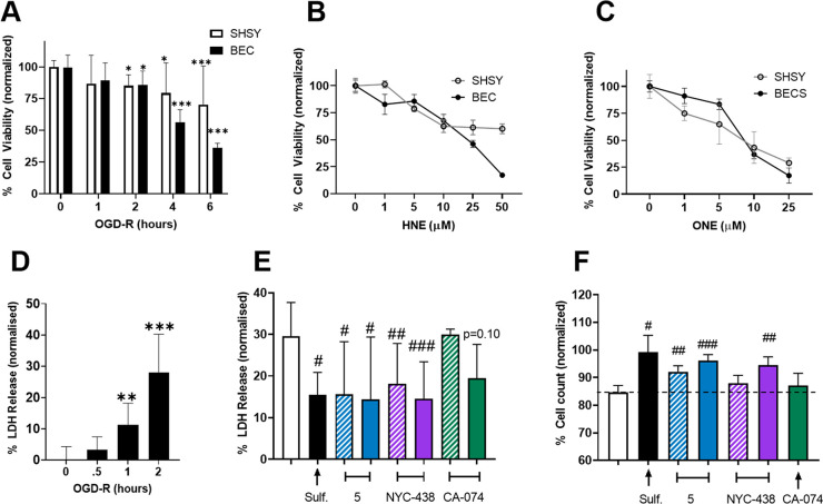
Figure 3.
BECs are sensitive to oxidative stress and are protected by calpain inhibitors. (A) Cell viability quantified by MTS of BECs and SH-SY5Y cell cultures subjected to 0, 1, 2, 4, or 6 h of OGD-R. (B and C) Cell viability quantified by MTS of BECs and SH-SY5Y cells after 24 h of treatment with the lipid peroxidation products HNE (B) or ONE (C). (D) Cell death quantified by LDH release of BECs following 0, 0.5, 1, or 2 h of OGD-R. (E) LDH release of BECs following 2 h of OGD-R with cotreatment of vehicle, sulforaphane (1 μM), or inhibitors (1 μM hashed; 10 μM solid) normalized to vehicle control (0%). (F) Cell count of BECs following 2 h of OGD-R and cotreatment of vehicle, sulforaphane (1 μM), or inhibitors (1 μM hashed;10 μM solid), normalized to in-plate sulforaphane (100%) with significance tested relative to vehicle control. Data represent mean ± SD of at least n = 3 in 3 separate isolations analyzed by one-way ANOVA with Dunnett’s or Tukey’s multicomparison: *, p < 0.05; **, p < 0.01; and ***, p < 0.001, versus no OGD-R control; #, p < 0.05; ##, p < 0.01; and ###, p < 0.001, versus vehicle-treated.

