Figure 2.
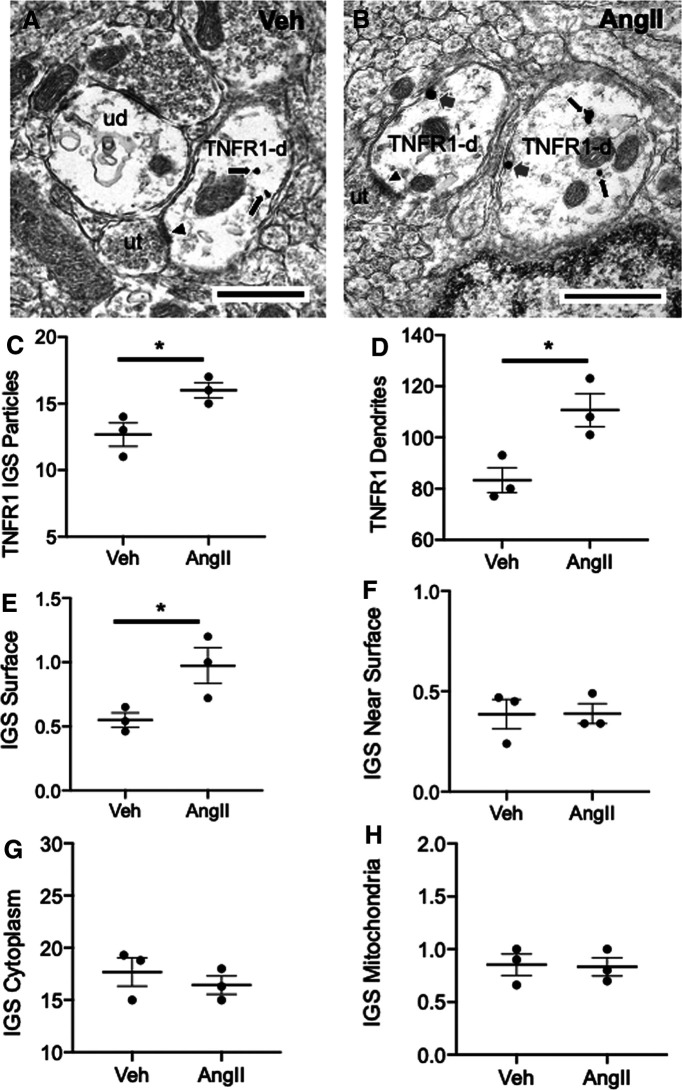
Chronic infusion of slow-pressor AngII is associated with increased TNFR1 labeling and elevated plasma membrane-affiliated TNFR1 in PVN neurons. A, B, Electron micrographs illustrating TNFR1 IGS labeling in dendrites (TNFR1-d) of PVN neurons from vehicle (Veh)-infused (A) and AngII-infused (B) mice. Immunoreactivity for TNFR1 is seen in the cytoplasm (black arrows) in both Veh-infused (A) and AngII-infused (B) mice. In addition, TNFR1 labeling is also present on the plasma membrane (gray arrows) of dendrites in mice receiving AngII. Some TNFR1 dendritic profiles receive asymmetric-type excitatory synapses (arrowheads) from unlabeled axon terminals (ut). C, Quantitatively, compared with Veh-treated mice, animals receiving AngII had a significantly greater number of IGS particles for TNFR1 in dendritic profiles. D, In addition, the number of TNFR1-labeled dendritic profiles was higher in AngII-infused mice compared with Veh-treated animals. There were also differences in the subcellular distribution of IGS particles for TNFR1 in AngII- and Veh-administered mice. E, In particular, the density of TNFR1 on the dendritic plasma membrane was greater in mice infused with AngII compared with Veh. F–H, There were no significant differences in the densities of TNFR1 labeling near the plasma membrane (F), in the cytoplasm (G), or affiliated with mitochondria (H) in either treatment group. *p < 0.05 AngII compared to Veh. Scale bars, 500 nm.

