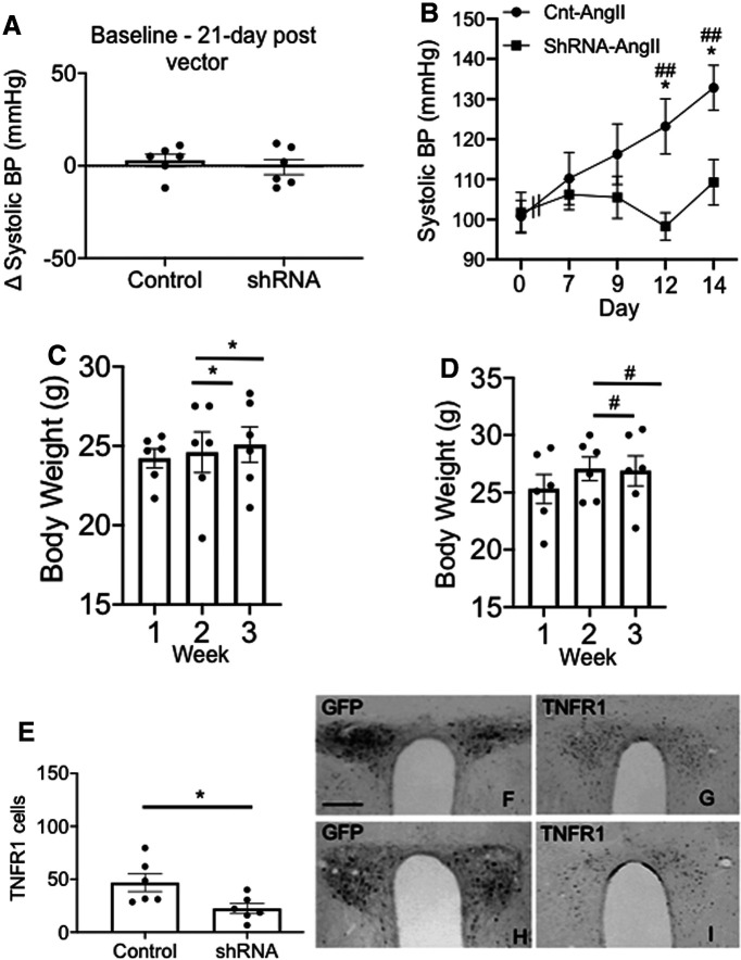
Figure 9.
Spatial-temporal knockdown of TNFR1 in PVN neurons is associated with an inhibition of the hypertensive response to slow-pressor AngII infusion. A, Mice receiving bilateral microinjection of control or shRNA vectors did not differ in systemic systolic blood pressure 21 d postinjection versus preinjection. B, There was a significant difference in the hypertensive response to AngII in the control and test vector-treated groups (*p < 0.02, control vs shRNA on day 12 and day 14; #p < 0.02, ##p < 0.005, control day 0 vs days 12 and 14). Mice microinjected with the control vector showed a significant increase in blood pressure, whereas mice receiving the shRNA vector did not demonstrate any treatment-dependent elevation in blood pressure over the 14 d infusion period. C, D, There were no significant differences in body weight gain postinjection (*p < 0.05, control weeks 1–3; #p < 0.05, shRNA weeks 1–3). E, There was a significant reduction in TNFR1 expression in mice microinjected with the TNFR1 shRNA (*p < 0.05). F, G, Micrographs illustrating bilateral expression of the reporter protein GFP (F) and TNFR1 (G) in the PVN are shown after bilateral microinjection of control vector. H, I, Bilateral expression of GFP (H) and TNFR1 (I) is also shown after TNFR1 shRNA microinjection. Scale bar, 0.5 mm.

