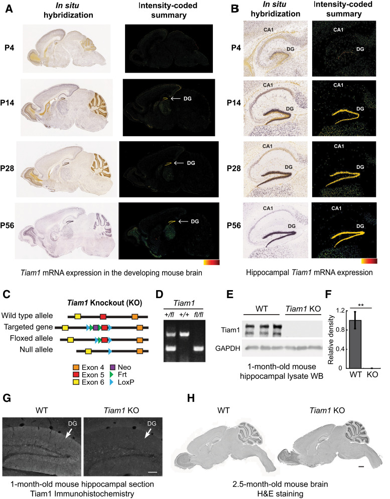
Figure 1.
Generation and characterization of Tiam1 KO mice. A, In situ hybridization (ISH) images (left) of Tiam1 mRNA in sagittal brain sections from different aged mice demonstrating the developmental expression of Tiam1. Intensity-coded summary images (right) show low (yellow) to high (red) Tiam1 expression. P, Postnatal day. Image credit: Allen Institute. B, Enlarged view of ISH (left) and intensity-coded summary (right) images from A showing Tiam1 expression in the hippocampus, where it is highly enriched in the DG. Image credit: Allen Institute. C, To target the murine Tiam1 gene, two loxP sites were inserted into a region flanking exon 5 and an internal Frt-flanked neomycin (Neo) cassette was added as a selection marker. The floxed allele (Tiam1fl/fl) was generated after removing Neo via breeding with Flippase-expressing mice. Tiam1fl/fl mice were then crossed with Ella-Cre mice to delete Tiam1 globally from early embryos, creating Tiam1−/− mice. For all figures, mice are abbreviated as follows: WT, Tiam1−/− (Tiam1 KO or KO). D, PCR analysis of tail DNA prepared from Tiam1+/fl, Tiam1+/+, and Tiam1fl/fl mice. E, Representative immunoblots of hippocampal lysates from 1-month-old WT and Tiam1 KO mice probed with antibodies against Tiam1 and GAPDH (loading control) demonstrating loss of Tiam1. F, Quantification of immunoblots from E (t(4) = 5.687, p = 0.005, unpaired t test, N = 3 mice per genotype). G, Representative immunohistochemistry images of coronal hippocampal sections from 1-month-old WT and Tiam1 KO mice showing loss of Tiam1 staining from the DG of Tiam1 KO mice. Scale bar, 100 µm. H, H&E staining of sagittal brain section from 2.5-month-old WT and Tiam1 KO mice demonstrating no gross changes in brain structure as a result of Tiam1 loss. Scale bar, 1000 µm.

