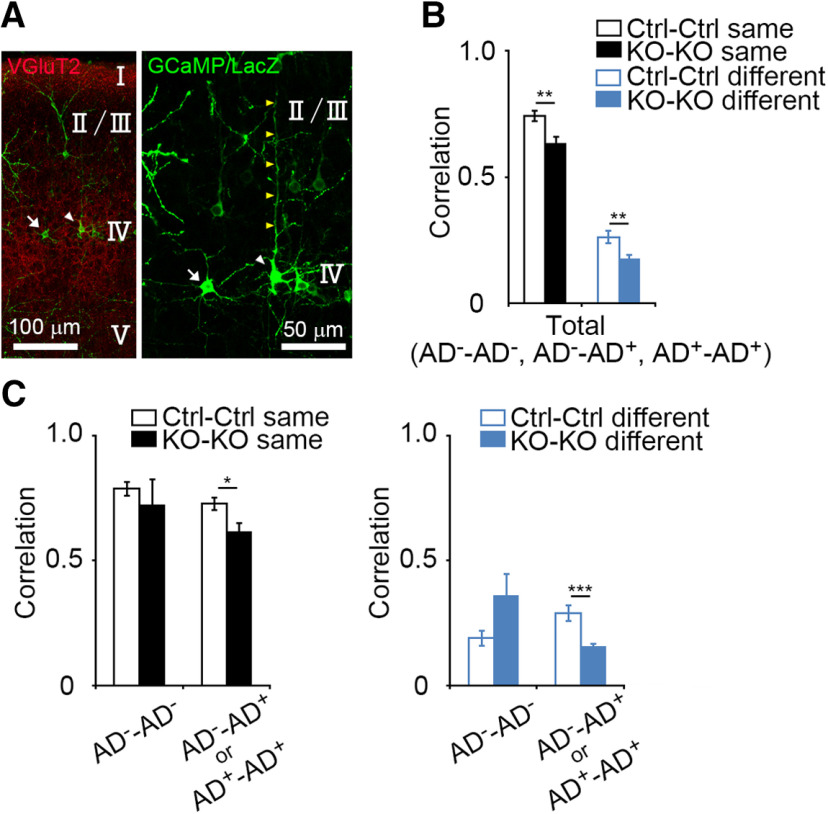
Figure 6.
Lower activity correlation in apical dendrite-possessing GluN1KO neurons. A, Representative images of an L4 neuron lacking an apical dendrite (AD−: a mature SS neuron), and an L4 neuron with an apical dendrite (AD+: an SP neuron or immature SS neuron) in the coronal section at P6. Arrows: an AD− neuron; white arrowheads: an AD+ neuron; yellow arrowheads: an apical dendrite. The right panel is higher magnification image of the left panel. B, Comparisons of correlation values between control neuron pairs and GluN1KO neuron pairs (control same vs GluN1KO same, p = 0.003; control different vs GluN1KO different, p = 0.006). In these analyses, cells whose cell-types were not clear were excluded. C, Relationship between activity correlation and apical dendrite morphology. The activity correlation of cell pairs that contain at least one AD+ cell was lower in GluN1KO than in control (same barrel, p = 0.012; different barrels, p < 0.001). Error bars indicate SE.

