Figure 1.

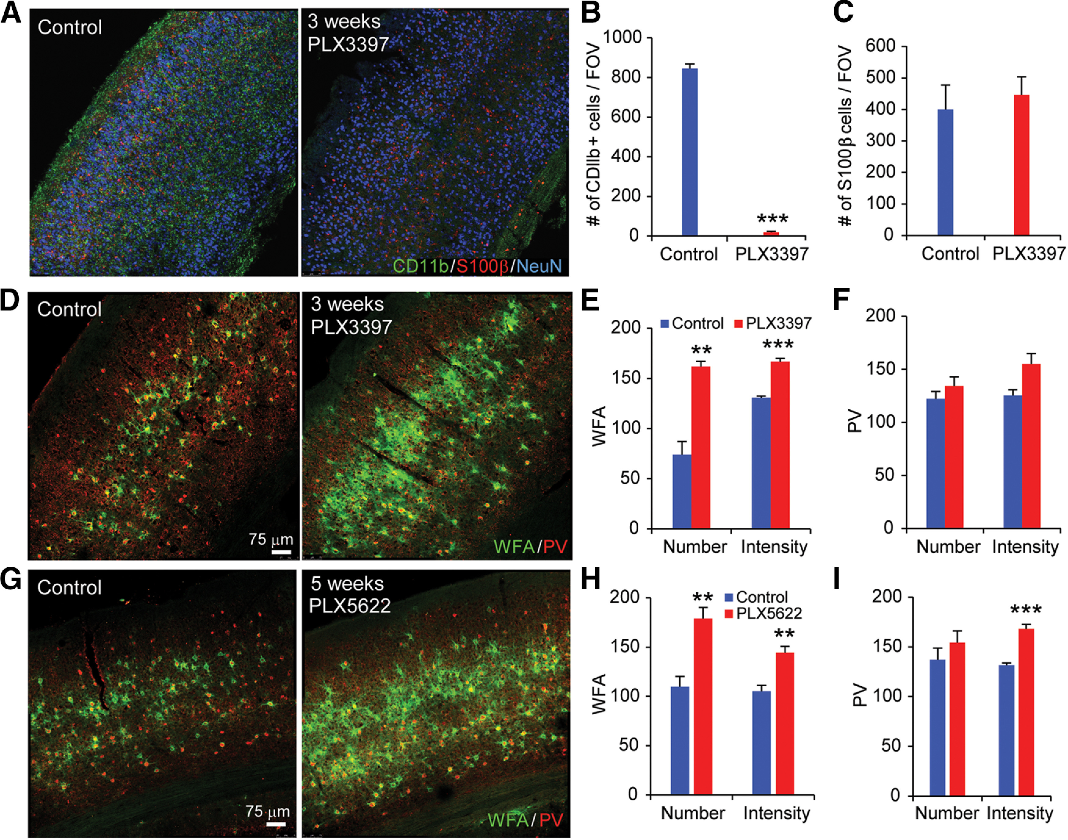
Microglial depletion by treatment with CSF1R inhibitors PLX3397 and PLX5622 robustly enhances PNNs. A, WT C57BL6/J mice treated for 3 weeks with control diet, or PLX3397 (290 ppm in chow) or PLX5622 (1200 ppm in chow). CD11b, S100β, and NeuN staining in visual cortex sections is shown for control (left) and PLX3397 treatment of 3 weeks (right). B, Numbers of CD11b+ cells/FOV are plotted with control and PLX3397 treatment. Microglia are reduced by ∼95% with this PLX3397 treatment (p = 0.001). C, Quantification of the number of S100β+ cells/FOV for control and PLX3397 conditions showing that astrocyte intensities were unaffected by PLX3397. D, WFA and PV staining is shown for control condition (left) and for PLX3397 treatment (right). E, Quantification shows that the numbers and intensity of WFA-positive PNNs per FOV significantly increase following 3-week-long PLX3397 treatment (p = 0.003 and p = 5.56 × 10⁻⁴). F, The PLX3397 treatment does not modulate the number and intensity of PV neurons (p = 0.335 and p = 0.056). G, WFA and PV staining following 5-week-long PLX5622 treatment. WFA and PV numbers and intensities for each condition are plotted in H and I. WFA is significantly increased with PLX5622 treatment (p = 0.002 for WFA number; and p = 0.002 for WFA intensity). Although the numbers of PV neurons are not affected (p = 0.359), PV staining intensity is significantly increased with PLX5622 treatment (p = 2.93 × 10⁻⁴). Data are mean ± SEM (samples from at least 3 mice/group). **p ≤ 0.01; ***p ≤ 0.001; two- tailed unpaired Student's t test.
