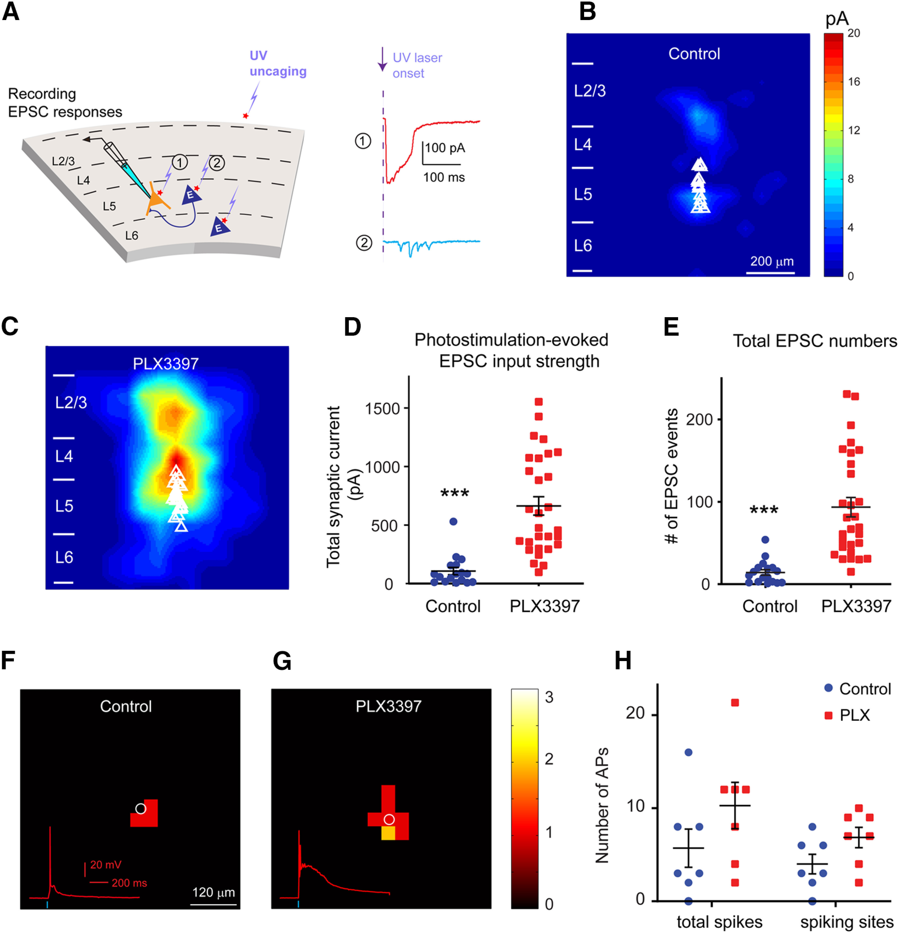
Figure 3.

LSPS mapping in brain slices reveals enhanced local excitatory connections to excitatory pyramidal neurons in adult mouse visual cortex following 3-week-long PLX3397 treatment. A, Schematic of LSPS. During the experiment, a layer 5 pyramidal neuron (left) is recorded by patch clamping while stimulating its surrounding sites by short-duration UV glutamate uncaging to generate action potentials from potentially connected presynaptic neurons. Right, Example uncaging responses. Direct uncaging responses (1, red) are excluded from synaptic input analysis, and synaptically mediated EPSC responses (2, cyan) are plotted to show latency and amplitude characteristics of synaptic inputs from presynaptic neuronal spiking. B, C, The averaged photostimulation-evoked EPSC input maps from visual cortex in control mice (n = 17 cells from 8 mice) and PLX3397 treatment mice (n = 29 cells from 10 mice), respectively. D, Average strengths of summed EPSC inputs in mice treated with PLX3397 (n = 29 cells) increase significantly compared with control cells (n = 17 cells) in adult mice (p = 1.48 × 10⁻⁶). E, The numbers of total EPSC input events per cell for each group differ significantly (control vs PLX3397, p = 3.97 × 10⁻⁷). Data are mean ± SEM. ***p < 0.001 (Mann–Whitney U tests). F–H, Spiking excitation profiles of excitatory neurons measured by glutamate uncaging show a trend of increase in excitability for the recorded cells in PLX3397-treated cortex (n = 7 cells from 4 mice), compared with controls (n = 7 cells from 4 mice; p = 0.17 for total spikes; p = 0.085 for spiking sites, Mann–Whitney U test).
