Figure 2.
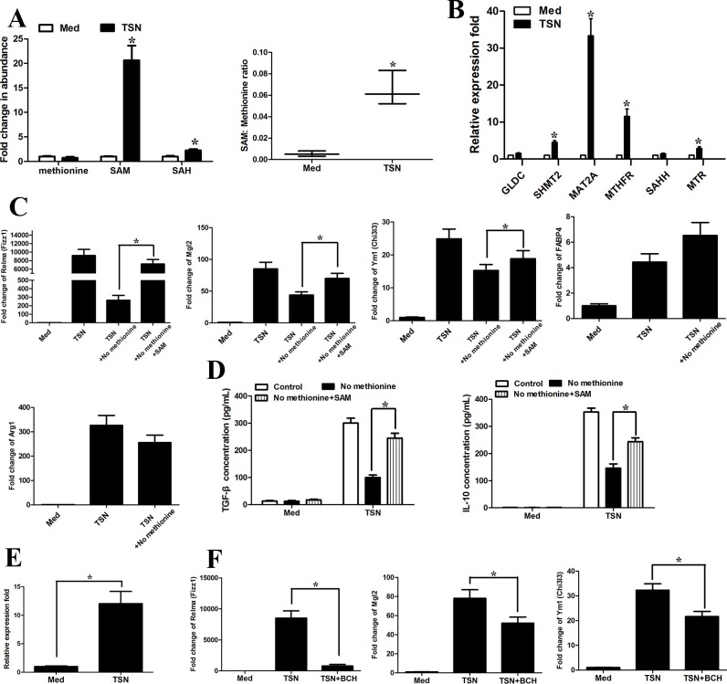
Methionine mediates tumor-induced M2 phenotype of monocytes. CD14+ cells were isolated from peripheral blood of healthy donors. CD14+ cells were left untreated or treated with TSN from MGC803 cells for 20 hours. (A) (Left) LC-MS was used to determine the abundance of methionine cycle metabolites in CD14+ cells purified from tumor tissues 48 hours after methionine starvation. Values were normalized to that of CD14+ cells in the absence of TSN. (Right) Ratio of SAM to methionine levels in CD14+ cells. (B) The expression levels of methionine metabolism-related gene with qRT-PCR. (C, D) Levels of the M2-associated genes Relma, Mgl2, Ym1, Fabp4, Arg1 by qPCR (C) and IL-10 and TGF-β production (D) by ELISA. (E) qRT-PCR analysis of the expression of amino acid transporter SLC7A5 on TSN treatment. (F) CD14+ cells were pretreated with DMSO or system L transporters (BCH, 5 mM) for 1 hour and then were exposed to medium (Med) or TSN for 20 hours. qRT-PCR analysis of the M2-associated genes Mgl2, Ym1 (Chi3I3) and Relma (Fizz1). Data are presented as the mean±SD; *p<0.05. N=3 biologically independent experiments. SAM, S-adenosylmethionine; TSN, tumor culture supernatant.

