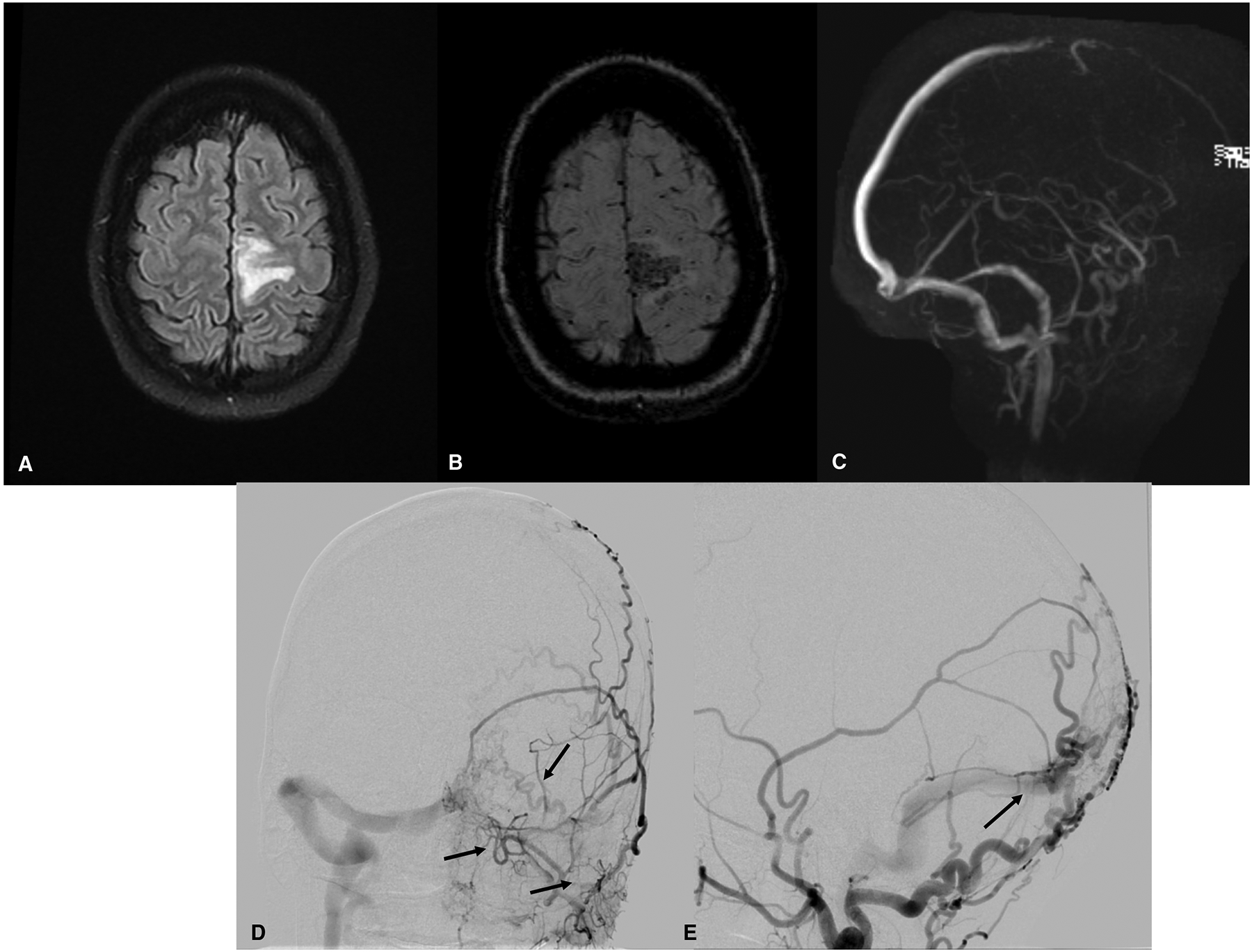
Figure 1.

Brain and vascular imaging findings in a 34-year primigravida at 14 weeks gestation, presenting with right leg weakness for hours, followed by a focal motor seizure of the right leg and arm that progressed to a generalized tonic-clonic seizure. (A) Brain magnetic resonance imaging (MRI, fluid-attenuated inversion recovery (FLAIR) sequence) shows cerebral edema in the left frontal lobe. (B) Susceptibility-weighted MRI shows hemorrhage in the same region. (C) Magnetic resonance venography shows thrombo-occlusion in the anterior portion of the superior sagittal sinus and decreased flow-related enhancement of cortical veins overlying the left hemisphere. The patient was diagnosed with cerebral venous sinus and cortical vein thrombosis. She received therapeutic anticoagulation with low-molecular-weight heparin. A subsequent digital subtraction angiogram of the brain revealed a dural arterial-venous fistula of the brain, with extensive arterial supply from the bilateral occipital arteries and middle meningeal arteries (D, E). Because the patient was clinically stable, treatment of the vascular malformation was postponed until after delivery.
