Fig. 6.

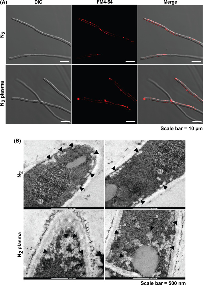
Visualization of secretory vesicles in the fungal hyphae after plasma treatment.
A. The vesicles in the fungal hyphae stained with FM4‐64 after 24 h. DIC denotes differential interference contrast microscopy.
B. The internal ultrastructure of the fungal hyphae after 24 h. The left and right panels show the tip and middle section of the fungal hyphae, respectively. The arrows indicate secretory vesicles.
