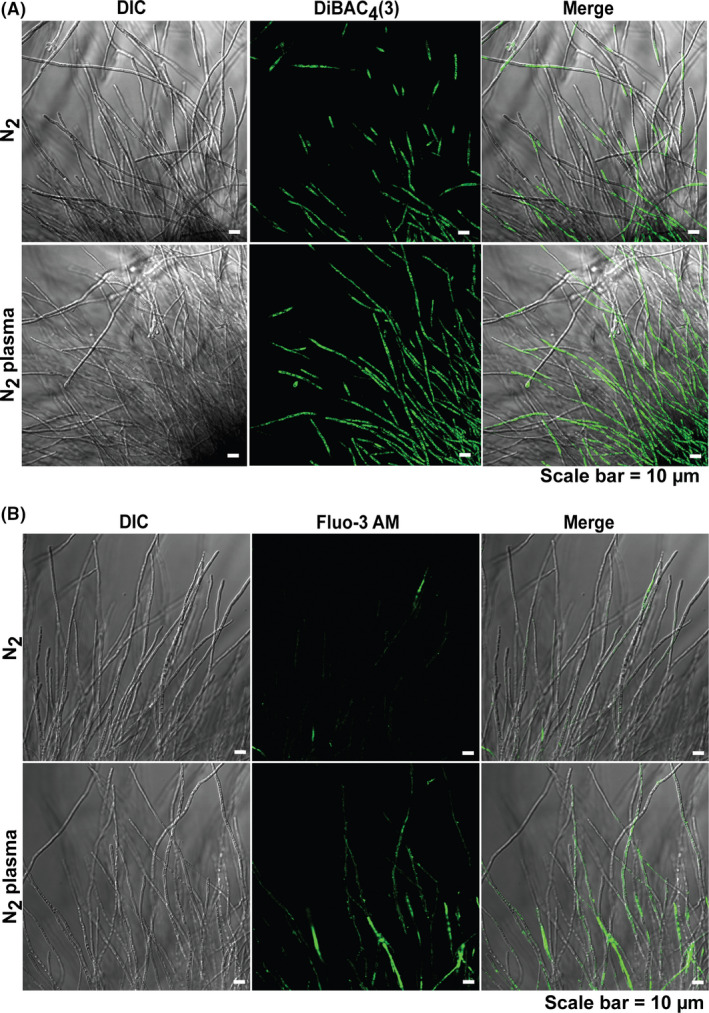
Fig. 7.

Analysis of membrane potential and intracellular Ca2+ levels in the fungal hyphae.
A. Fungal hyphae labelled with DiBAC4(3). Fluorescence occurred upon membrane depolarization.
B. Intracellular Ca2+ in fungal hyphae after 24 h following staining with Fluo‐3‐AM.
