
Figure 1.
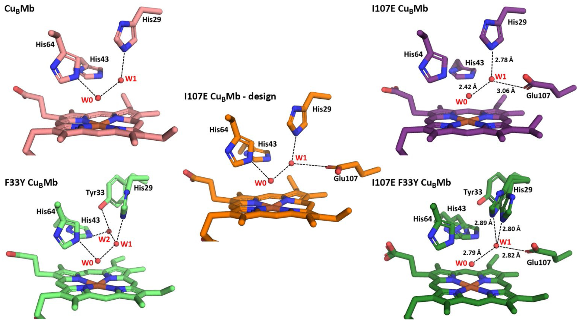
Structures of CuBMb variants. (left) Crystal structures of CuBMb and F33Y-CuBMb, highlighting water-mediated hydrogen-bonding network in the active site. (center) Computational model of I107E-CuBMb based on crystal structure of CuBMb and I107E containing Mb variant from PDB 3M38. (right) Crystal structures of I107E-CuBMb and I107E/F33Y-CuBMb.