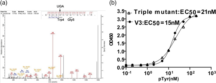
FIGURE 3.

Decoding of the opal stop codon UAG and its role in a functional SH2 variant. (a) The V3 variant was expressed and purified. The sample was trypsin digested and submitted for mass spectrometry assay. (b) The EC50 assay of the V3 and triple mutant variants binding to the phosphotyrosine peptide (EPQpYEEIPIYL)
