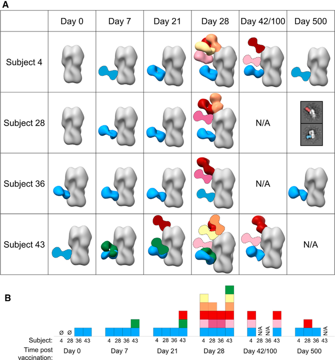
Figure 4. Polyclonal antibodies elicited by H5N1 vaccination decorate the stem, head, and vestigial esterase domains of HA.
(A) Matrix of negative stain EM reconstructions of pAbs in complex with recombinant H5 HA (A/Indonesia/5/2005) from each subject at all time points listed. 3D reconstructions of polyclonal immune complexes are shown for most time points. Due to limited particle representation, Fab graphics with dashed outlines are predicted placements. For samples with immune complexes in low abundance, example 2D class averages with labels are shown.
(B) Summary of epitopes targeted by pAbs. Each square represents a Fab specificity from the corresponding subject and time point. Stem specificities, blue and green; RBS-proximal and lateral patch specificities, red, orange, and yellow; vestigial esterase and mid-lateral head specificities, light pink and dark pink.
See also Figure S4.

