
Figure 4.
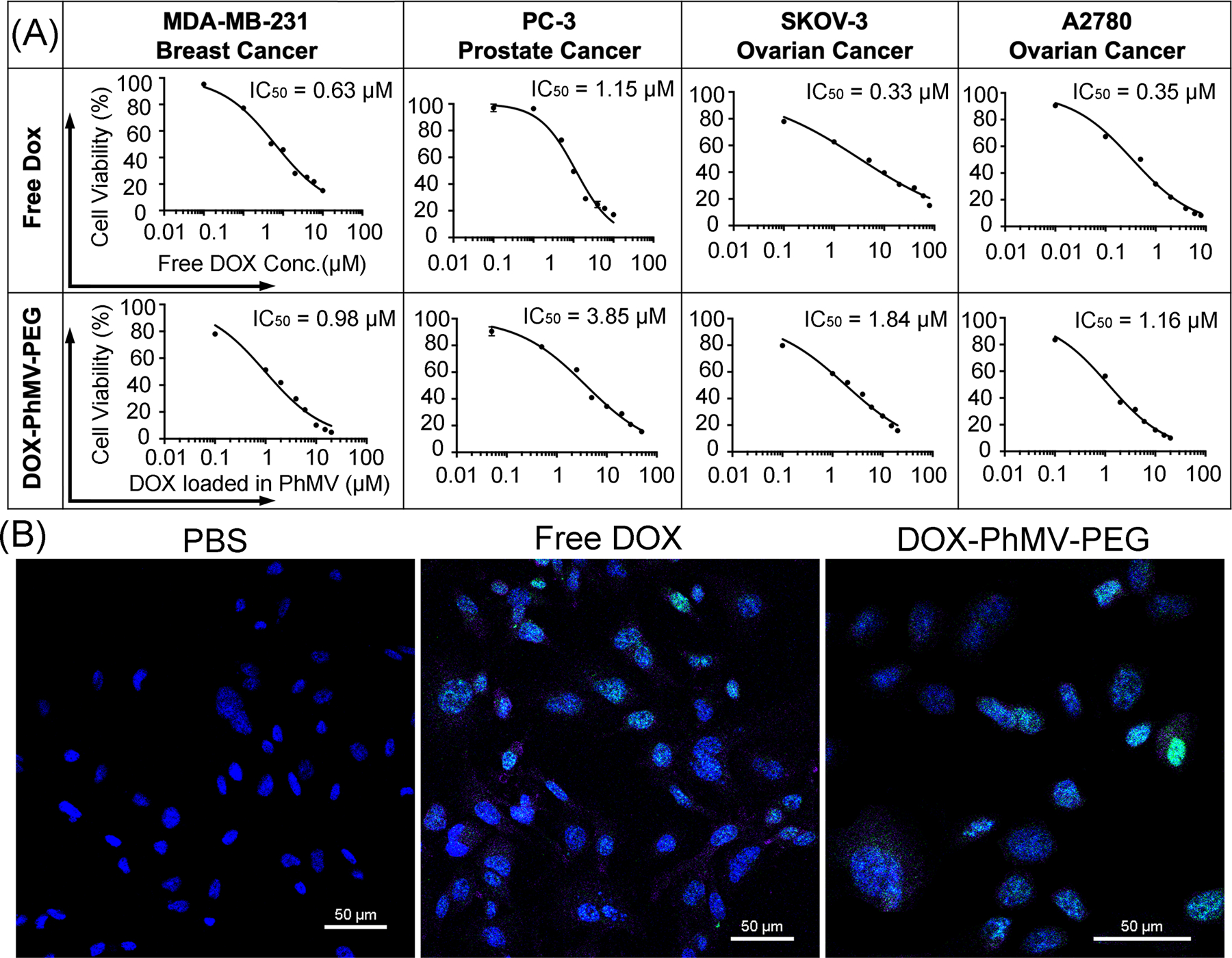
Cytotoxicity and DNA damage caused by DOX-PhMV-PEG particles. (A) The 50% inhibitory concentration (IC50) of free doxorubicin and DOX-PhMV-PEG particles against a panel of cancer cells. (B) Confocal images of MDA-MB-231 breast cancer cell reveal H2A.X phosphorylation (green) in nuclei (blue) following treatment with DOX-PhMV-PEG particles compared to free doxorubicin and PBS (scale bar = 50 µm).