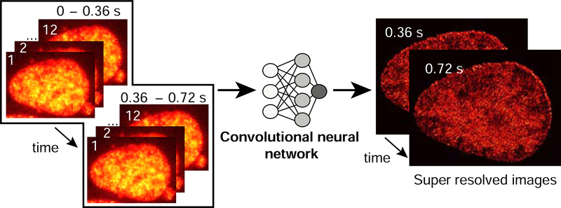
Figure 3. Deep-PALM uses a convolutional neural network to super-resolved images.
Photoactivated localization microscopy images of U2OS nuclei expressing H2B-PATagRFP are input to a trained deep convolutional neural network (CNN). The predictions from multiple input frames (30 ms/frame) are summed to construct a super-resolved image of chromatin in vivo with a final frame interval of 360 ms.

