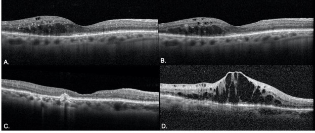
Fig 5. Example of DME disease progression on OCT from pre- to follow-up visit in the delayed and non-delayed groups.

A-B. A patient with DME presented (A) for pre-lockdown visit with stable disease. The patient returned for 10 week follow-up (B) with a stable degree of intraretinal fluid. C-D. Another patient presented for a pre-lockdown visit with stable disease (C) with a plan for follow up in 8 weeks. Follow up was delayed by 6 weeks and OCT revealed significant accumulation of intraretinal and subretinal fluid.
