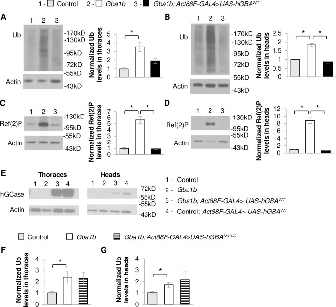
Fig 10. Muscle expression of human WT GBA suppresses protein aggregation in Gba1b mutants.
(A-E) Using the flight muscle specific driver, Act88F-GAL4, wildtype (WT) human GBA (hGBAWT) was expressed in Gba1b mutant and WT revertant controls. Homogenates were prepared from fly heads and thoraces using 1% Triton X-100 lysis buffer. Western blot analysis was performed on the Triton X-100 insoluble proteins using antibodies to ubiquitin (Ub) and Actin, and on the soluble fractions using antibodies to Ref(2)P, Actin, and human glucocerebrosidase (hGCase). Representative images and quantification of ubiquitin in (A) thoraces (One-way ANOVA: F(2,6) = 19.949, p = 0.002) and (B) heads (F(2,6) = 24.854, p = 0.001) and Ref(2)P in (C) thoraces (F(2,6) = 10.609, p = 0.011) and (D) heads (F(2,6) = 23.297, p = 0.001) of Gba1b mutant flies with and without muscle expression of hGBAWT are shown. Results are normalized to Actin and control. (E) Antibody detecting hGCase in the thoraces and heads of control and Gba1b mutant flies with and without muscle expression of hGBAWT. (F,G) Using Act88F-GAL4, mutant human GBA (hGBAN370S) was expressed in Gba1b mutant and controls. Western blot analysis was performed on the Triton X-100 insoluble proteins using antibodies to ubiquitin (Ub) and Actin. Quantification of ubiquitin in (F) thoraces (F(2,6) = 5.190, p = 0.049) and (G) heads (F(2,6) = 7.250, p = 0.025) of Gba1b mutant flies with and without muscle expression of hGBAN370S are shown. Results are normalized to Actin and control. (Representative images are found in S7 Fig). At least 3 independent experiments were performed. Error bars represent SEM. *p < 0.05 by Student t-test.

