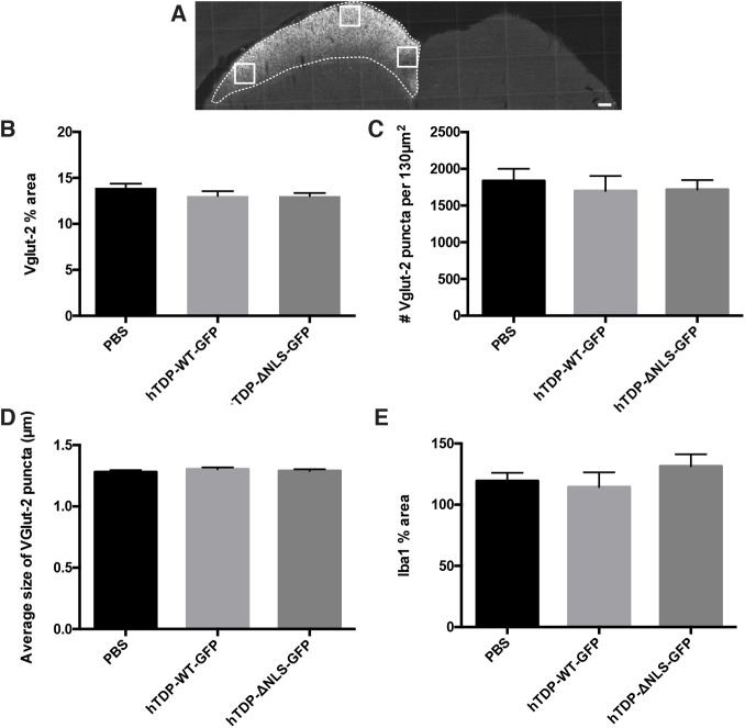
Fig. 8.
TDP-43 alterations do not cause RGC pre-synaptic changes. (A) Representative image of coronally sectioned brain showing RGC terminals within the SC following intraocular injection with choleratoxin subunit b (CTB). White boxes indicate ROIs used for Vglut2 analysis, and dotted white lines indicate the edge of CTB labelling, used for Iba1 analysis. Mice were injected with vehicle (PBS), hTDP-WT-GFP or hTDP-ΔNLS-GFP. (B-D) Quantitation of segmented puncta pooled from three ROIs immunolabelled with Vglut2 yielded measurements of percentage area (B), average number (C) and average size (D) of Vglut2-positive synaptic boutons. (E) Quantitation of segmented microglia immunolabelled with Iba-1 within CTB-labelled SC yielded the percentage area occupied by Iba1-positive microglia, when normalized to the same ROI on the contralateral SC. Values are mean±s.e.m., n=5 per treatment group. Data were analysed with one-way ANOVAs with Tukey post hoc tests, showing no significant differences. Scale bar: 150 µm.

