
Fig. 2. DLSμR captures actin-dependent matrix dynamics and cell-mediated remodeling of viscoelasticity.
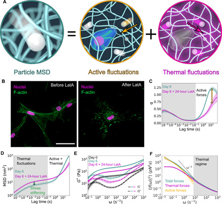
(A) Schematic of the contributions to particle motion from thermal and active fluctuations. (B) Confocal microscopy of HMFs cultured in col/rBM for 6 days before and after depolymerization of the F-actin cytoskeleton with latrunculin A (LatA). F-actin fibers (green false color) were stained with TRITC-phalloidin. Nuclei (magenta false color) were counterstained with DAPI. Scale bar, 50 μm. (C) Power-law scaling of the MSD, α, before and after latrunculin A treatment. Shading designates regimes where α is independent (gray) or dependent (white) on F-actin polymerization. (D) Particle MSD. Shaded regions denote regimes governed by either thermal fluctuations (gray) or a superposition of thermal and active fluctuations (white). (E) Shear modulus, G*, of the ECM as a function of angular frequency ω at indicated culture time points. Solid and dashed lines denote the storage modulus G′ and loss modulus G″, respectively. (F) Force fluctuations at different frequencies ω on day 6. The shaded region designates where thermal forces dominate over active forces. The dashed black line shows the high-frequency scaling of step-like motor forces, 〈∣f(ω)∣2〉 ∼ ω−2. Curves represent the arithmetic (C, E, and F) mean or geometric (D) mean among biological replicates (n = 7). Error bands represent 68% confidence intervals of the mean.