
Fig. 2. FACS selection for variants of S with high or low binding signal to ACE2.
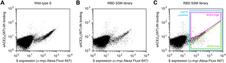
(A) Flow cytometry analysis of Expi293F cells expressing full-length S of SARS-CoV-2 with an N-terminal c-myc tag. Staining for the myc-epitope is on the x axis, while the detection of bound sACE22-8h (2.5 nM) is on the y axis. S plasmid was diluted 1500-fold by weight with carrier DNA so that cells typically express no more than one coding variant; under these conditions, most cells are negative. (B) Flow cytometry of cells transfected with the RBD single site-saturation mutagenesis (SSM) library shows cells expressing S variants with reduced sACE22-8h binding. (C) Gating strategy for FACS. S-expressing cells positive for the c-myc epitope were gated (blue), and the highest (“ACE2-high”) and lowest (“ACE2-low”) 20% of cells with bound sACE22-8h relative to myc-S expression were collected.