
Fig. 6. Polyamines promote the metabolism of TAMCs under conditions of pHe.
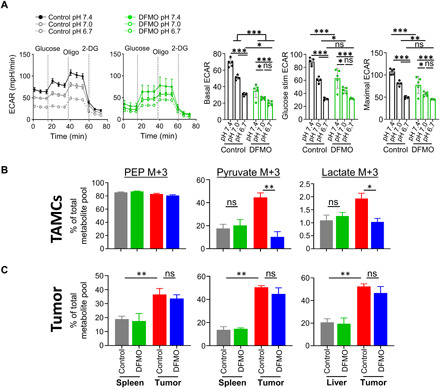
(A) TAMCs were generated in the context of DFMO, and after 6 days of differentiation, cells were lifted, adhered using Cell-Tak adhesive, and cultured in decreasing pH medium. Immediately following addition of acidic medium, Seahorse extracellular flux analysis was performed to determine glycolytic metabolism. (B) DFMO-treated animals had TAMCs isolated, and ex vivo 13C-glucose flux was performed. (C) In vivo 13C-glucose flux was performed by infusion of 13C-glucose (100 mg/ml) over a 2.5-hour period. After infusion, tumor, adjacent parenchyma, and liver were flash-frozen for bulk metabolite analysis. (A) n = 5 per group, representative of two independent experiments. (B) Each sample is n = 10 mice pooled per sample, with three pooled samples submitted in triplicate; data are representative of two independent experiments. (C) n = 3 mice were individually analyzed per group. Statistics were calculated via unpaired Student’s t tests: *P < 0.05, **P < 0.01, and ***P < 0.001. 2-DG, 2-deoxyglucose; PEP, phosphoenolpyruvate.