
Fig. 4. Effect of ICIs and IDO-1 inhibitors in traditional assays and the tumor-on-a-chip.
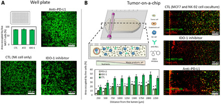
(A) The potential effect of atezolizumab (anti–PD-L1 antibody) and epacadostat (IDO-1 inhibitor) was evaluated in traditional well plates. A confluent monolayer of MCF7 was seeded on 96-well plates, and NK-92 cells were added 24 hours later (9 MCF7:1 NK ratio) with/without the IDO-1 inhibitor or the PD-L1 antibody. Confocal images showed no significant improvement on NK-92 cell cytotoxic potential. (B) Similar experiment evaluating epacadostat and atezolizumab in the tumor-on-a-chip microdevice. Confocal images demonstrated that both the PD-L1 antibody and IDO-1 inhibitor increased MCF7 cell necrosis. Dead cells (shown in red) concentrated in the vicinities of the lumen, whereas live MCF7 cells (shown in green) remained present in the farthest (distal) region. Asterisk denotes P value of <0.05; graphs show means ± SD.