
Fig. 1. The expression and distribution of RLN receptor RXFP1 in desmoplastic tumors.
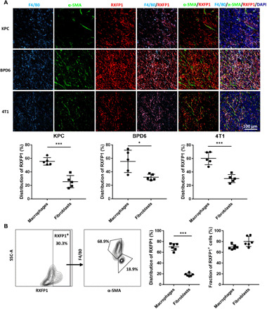
(A) The distribution of RXFP1 in three different desmoplastic tumors including KPC pancreatic cancer, BPD6 melanoma, and 4T1 breast cancer (n = 5 samples per group). Macrophages were stained with anti-F4/80 in cyan, and fibroblasts in the tumor tissue were stained with anti–α-SMA (green). RXFP1 was stained in red with the anti-RXFP1 antibody. Cell nuclei were stained with DAPI in blue. (B) The distribution of RXFP1 in KPC pancreatic tumor further evaluated by flow cytometry (n = 6). Statistical significance was calculated using t test. *P < 0.05, ***P < 0.001.