
Fig. 4. RLN gene delivery caused macrophage-involved fibrosis regression and T cell infiltration.
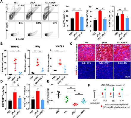
(A) Cotreatment of pRLN and CEL decreased the amount of F4/80+CD11b+ total macrophages and F4/80+CD206+ macrophages in the tumor tissue (n = 3). (B) Relative expression of MMP13, IFNγ, and CXCL9 detected by RT-PCR after different treatments (n = 4). (C) Collagen expression and CD3+ T cell infiltration in the tumor (n = 4 samples per group). (D) Tumor infiltrated CD3+CD4+ and CD3+CD8+ T cells quantified with flow cytometry (n = 3). (E) Tumor weight in each group after mice were euthanized on day 28. (F) The dosing scheme for the treatment of CEL and pRLN (n = 5 to 6). Statistical significance was calculated using one-way ANOVA with multiple comparisons. *P < 0.05, **P < 0.01, and ***P < 0.001.