
Fig. 5. RLN gene delivery improved therapy response to PD-L1 blockade in KPC pancreatic tumor.
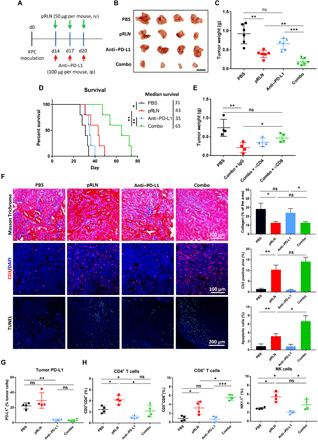
(A) Tumor inoculation and treatment scheme for the combination of pRLN and anti–PD-L1 therapy. (B) Representative images (scale bar, 1 cm) of tumors dissected from tumor bearing mice on day 28 and (C) weight of tumors quantified (n = 6). (D) Survival of mice after different treatments (n = 5). (E) Tumor weight of tumor bearing mice with the combination therapy and depletion of CD4+ and CD8+ T cells with antibodies (n = 4). (F) Collagen expression, CD3+ T cell infiltration, and apoptotic cell detection in the tumor tissue (n = 3 samples per group). Masson Trichrome assay was performed to detect collagen expression. IF staining and terminal deoxynucleotidyl transferase–mediated deoxyuridine triphosphate nick end labeling (TUNEL) assay were used to evaluate T cell (red) infiltration and apoptotic cells (green) in the tumor, respectively. Cell nuclei were stained with DAPI in blue. (G) PD-L1 expression on cells in the tumor (n = 4). (H) Immune cell infiltration in the tumor detected with flow cytometry (n = 4). Statistical significance was calculated in (C), (E), (F), (G), and (H) using one-way ANOVA with multiple comparisons, and statistical significance in (D) was calculated using log rank test. *P < 0.05, **P < 0.01, and ***P < 0.001.