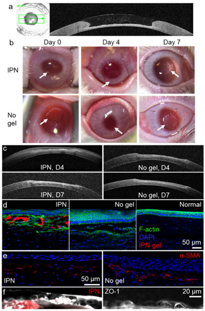
Figure 6.
Ex vivo and in vivo studies of the application of the IPN as a corneal defect filler. (a) OCT image shows the curvature restoration of the IPN on a rabbit corneal defect ex vivo. The IPN was added to the corneal defect when it is still in a liquid-like state. (b) Magnified photos of treated eyes on different days after treatment. The keratectomy area of the untreated group was more opaque than that treated with the IPN. The arrows indicate the edges of the keratectomy area. (c) OCT images of corneal defect recovery over 1 week. Immunofluorescence staining of regenerated anterior corneal tissues: (d) F-actin, (e) alpha smooth muscle actin, and (f) zonula occludens-1. Images on the same row share the same scale bar.

