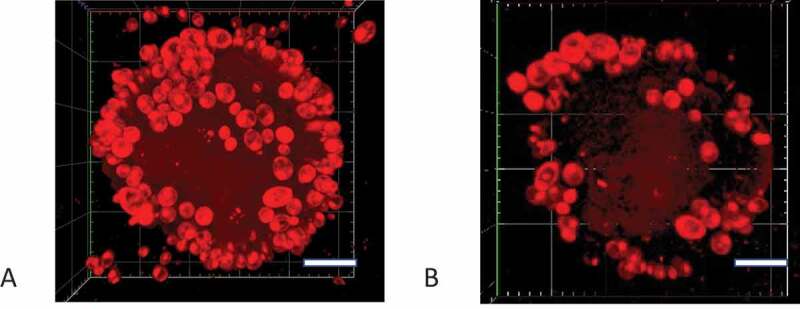
Figure 2.

Confocal microscopy Z-stack imaging to visualize C. albicans adhering to saliva-coated beads. The C. albicans SC5314 cells that attached to hydroxyapatite beads were stained with NR. Confocal images were captured with a Zeiss LSCM780 laser scanning microscope. Scale-bar: 20 µm. (A) Bead that had been incubated with C. albicans SC5314 cells at OD600nm = 1, (B) Bead that had been incubated with C. albicans SC5314 cells at OD600nm = 0.1
