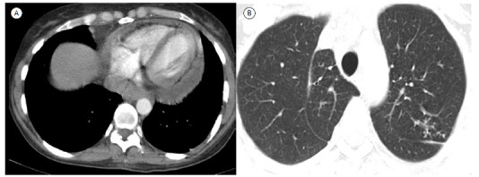
Figure 1. In A, an axial CT slice viewed at mediastinal window settings shows a fluid density collection involving the heart (pericardial effusion). In B, an axial CT slice viewed at parenchymal window settings shows nodular opacities in the upper lobe of the left lung, demonstrating the tree-in-bud pattern.

