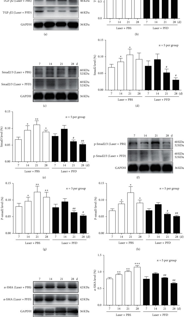
Figure 2.

Intravitreal injection of PFD attenuates the formation of fibrosis. Representative images of collagen I (red) staining of the retinal pigment epithelium-choroid-sclera flat mounts obtained from the three groups. Scale bar: 200 µm. Quantitative measurement of the fibrosis area. ∗∗P < 0.01 vs. Laser and Laser + PBS. #P < 0.05 vs. day 21.
