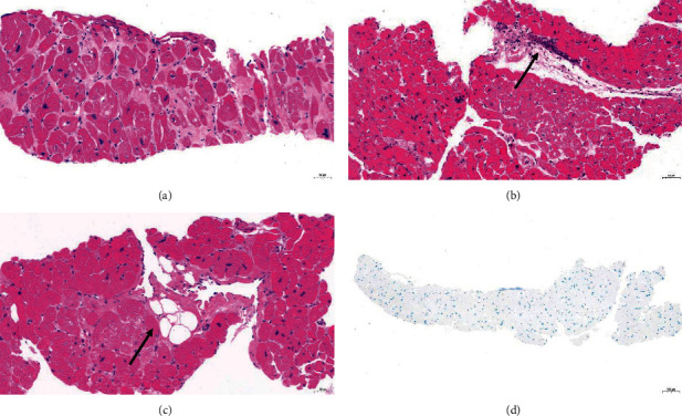
Figure 4.

Histopathology changes in HOCM specimens. (a) Myocytes hypertrophy and hyperchromatic nuclei with bizarre shapes. (b) Inflammatory cells infiltration. (c) Adipocyte infiltration. (d) No amyloidosis (H-E staining and Congo red staining 20x).
