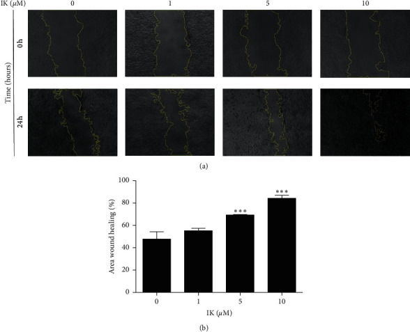
Figure 2.

Effects of IK on scratch wound closure of HaCaT cells in vitro. HaCaT cell monolayer was mechanically scratched and incubated with the indicated concentration of IK. HaCaT cell migration was monitored using a microscope at 0 and 24 h after scratch. (a) Images of HaCaT cells treated IK at 0 and 24 h at 10 × magnification. The yellow line shows the border of the wound. (b) Relative wound closure area of HaCaT cells according to IK concentration. The wound area was measured using Image J and calculated as the ratio of the scratch area at 24 h relative to 0 h was calculated. The results are presented as the means ± SD of three independent experiments ( ∗p < 0.05, ∗∗p < 0.01, and ∗∗∗p < 0.001 versus 0 μM group).
