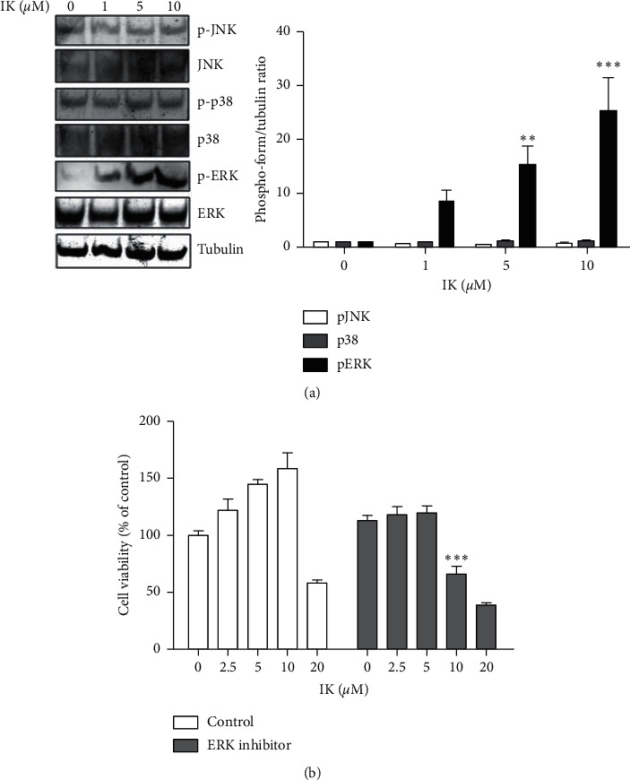
Figure 3.

IK enhances proliferation of keratinocytes through induction of ERK 1/2 pathway. (a) Western blot of HaCaT cells treated with varying concentrations of IK. HaCaT cells were treated with IK (0, 1, 5, and 10 μM) for 24 h. Whole-cell extracts were harvested and analyzed using the antibody against total JNK, ERK1/2, p38, phospho-JNK, ERK1/2, p38, and beta-tubulin was used as the loading control. (b) Viability of HaCaT cells compared with only IK treatments and cotreated with ERK 1/2 inhibitor, PD98059. HaCaT cells were seeded into a 96-well cell culture plate and treated with various concentrations of IK only or with ERK 1/2 inhibitor for 24 h. Cell viability was measured using an Ez-Cytox Kit. The results are presented as the means ± SD of three independent experiments ( ∗p < 0.05, ∗∗p < 0.01, and ∗∗∗p < 0.001 versus control group).
