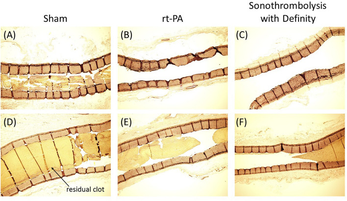
Figure 4.
Representative longitudinal histology images of residual clot burden in APAs with Verhoeff-Van Giessen (VVG) stain. Residual clot volume can be seen in vessels in the sham treatment arm (A, D) rt-PA only treatment arm (B, E) or the sonothrombolysis with Definity treatment arm. Images A–C show vessels for which the perfusion was graded as ≥ 2b at 120 min on the mTICI scale and images D-F show vessels graded < 2b at 120 min.

