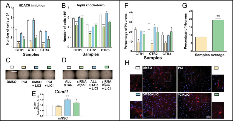
Fig. 3. Lithium rescues proliferation and differentiation capabilities in CdLS mouse NSCs through WNT activation.
A Analysis of the effects of PCI34051 and lithium exposure on the proliferation capabilities of the NSCs. White: NSCs treated with DMSO; yellow: NSCs treated with PCI34051; light blue: NSCs treated with DMSO + LiCl; dark green: NSCs treated with PCI34051 and LiCl. *** p < 0.001; $ p < 0.05; * DMSO vs PCI34051; $ PCI34051 vs PCI34051 + LiCl. B Analysis of the effects of Nipbl knockdown and lithium exposure on the proliferation capabilities of the NSCs. White: NSCs treated with AllStar; light green: NSCs treated with siRNA against Nipbl; light blue: NSCs treated with AllStar and LiCl; dark green: NSCs treated with siRNA against Nipbl and LiCl. *** and °°° p < 0.001; * AllStar vs siRNA against Nipbl; ° siRNA against Nipbl vs siRNA against Nipbl + LiCl. C Representative pictures of the neurospheres growing in the well during various treatments as in panel (A). Scale bar: 2 mm. D Representative pictures of the neurospheres growing in the well during various treatments as in panel (B). E Analysis of the effects of PCI34051 and lithium exposure on the gene expression of Ccnd1 in the NSCs. White: NSCs treated with DMSO; yellow: NSCs treated with PCI34051; light blue: NSCs treated with DMSO + LiCl; dark green: NSCs treated with PCI34051 and LiCl. ** p < 0.01; * p < 0.05; ** DMSO + LiCl vs PCI34051; * PCI34051 + LiCl vs PCI34051. F Analysis of the effects of PCI34051 and lithium on the differentiation capabilities of the NSCs. Cells were immunostained for neuron and nuclei detection. White: NSCs treated with DMSO; yellow: NSCs treated with PCI34051; light blue: NSCs treated with LiCl; dark green: NSCs treated with PCI34051 and LiCl. G Comparison of the effects of PCI34051 and PCI34051 + LiCl of NSCs differentiation. ** p < 0.01; * PCI34051 vs PCI34051 + LiCl. H Representative staining of the differentiated NSCs. Differentiated neurons were labeled with β-tubulin III antibody (red) and nuclei were labeled with DAPI (blue). Scale bar: 50 µm.

