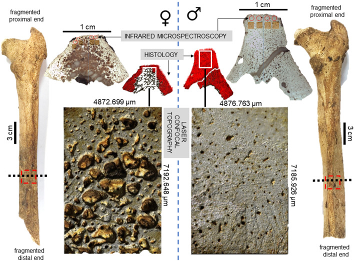
Figure 1.
Summary of methods and key findings in the present study. Posterior view of two right archaeological femora from Talasiu individuals estimated as female (♀, ID: Sk3.1) and male (♂, ID: Sk3.2) shows the sectioning location (black dashed line) and approximate sample removed (red dashed box). Methodological steps included synchrotron sourced infrared microspectroscopy to measure bone mineral composition, histomorphometry to estimate Haversian canal densities (red dots), and laser confocal scanning of bone topography to provide a qualitative illustration of intra-cortical bone porosity producing trabecularisation.

