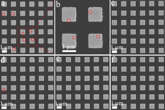
Fig. 5. Specificity of the digital nanopillar SERS platform for FGF-2 cytokine detection.

Representative SEM images of pillar array incubated with FGF-2 SERS nanotags in the presence of a, b FGF-2 (1031 aM), c G-CSF (1031 aM), d GM-CSF (1031 aM), e CX3CL1 (1031 aM), and f PBS. The red circles highlight the existence of SERS nanotags. Panel b is the magnified SEM image of the red-highlighted section in a. It is noted that nanofabrication debris on the sidewall of the pillars can also be seen. Data from one independent experiment.
