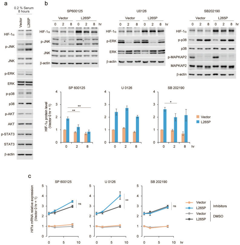
Figure 5.
JNK signalling is associated with HIF-1α protein level in MYD88 L265P-expressing p53−/−MEFs. (a) Vector and MYD88 L265P-expressing p53−/−MEFs were treated with growth medium containing 0.2% FBS for 8 h. (b) Vector and MYD88 L265P-expressing p53−/−MEFs were treated with the indicated MAPK inhibitors. Total cell lysates were analysed by immunoblotting (top). The quantification of HIF-1α signals was measured using ImageJ software and data were normalised to β-actin signals. The data represent untreated controls (vector), which were assigned a value of 1 (bottom). (c) Hif1a mRNA expression was measured by qPCR. The y-axis values are the relative fold change for gene transcripts. Each value was normalised to β-actin. (b,c) The data represent the mean ± s.d. using one-way ANOVA followed by Scheffe’s F test. *P < 0.05, **P < 0.01.

